Thursday 26 June 2014

Hydatid Cysts of The Liver


Hydatid Cysts of the Liver

In humans hydatid disease is caused by the larvae of a tapeworm called Echinococcus granulosus. This parasitic infection occurs worldwide and is endemic in some countries such as Australia and the Middle East, especially in sheep farming areas. Hydatid disease is a serious and potentially fatal condition, which may remain hidden in the body for many years.

How can humans become infected with this tapeworm?

Hydatid disease in humans is caused by contact with dog feces or dog hair infected with the tapeworm eggs, or contaminated vegetables. The eggs may stick to the animal’s hair or contaminate the vegetable garden. The eggs are highly resistant to the environment and can remain alive for months. Human infection does not occur from eating infected offal.  Hydatid disease is not contagious and is not passed by person-to-person contact.

Life cycle of a tapeworm

The life cycle of the tapeworm alternates between herbivores and carnivores—(typically sheep, foxes and dogs). Humans are an accidental intermediate host and become an end point in the tapeworm’s lifecycle.
hydatid life cycle
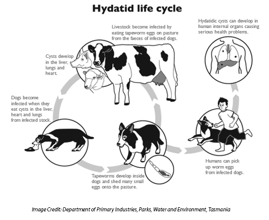
The sheep ingests the eggs which hatch in the sheep’s intestine and then travel to the liver where the hydatid cyst develops. When a dog eats the sheep’s organs containing the hydatid cyst, the dog becomes infected and passes eggs out in their feces. Cows and sheep become infected by eating the grass contaminated by dog feces. Other animals that may be infected include pigs, cattle, goats, horses, camels, wombats, wallabies and kangaroos. The grazing animal eats dog, fox or dingo feces infected with tapeworm eggs.  Eventually the animal’s organs (such as the liver, brain or lungs) grow watery sacs called hydatid cysts. These cysts contain tapeworm heads and a mature cyst may contain several million such heads.

Symptoms of Hydatid Disease in Humans

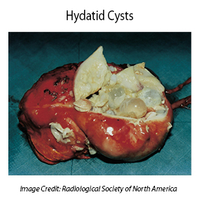
The symptoms of hydatid disease vary according to which body organs are infected. The most commonly infected organ is the liver, but the brain, kidneys and lungs are sometimes affected. The slow growing cysts are localized in the liver (in 75% of cases), the lungs (in 5-15% of cases) and other organs in the body such as the spleen, brain, heart and kidneys (in 10-20% of cases). The cysts are usually filled with a clear fluid called hydatid fluid and are spherical and usually consist of one compartment.
Symptoms can occur a long time after infection and sometimes many years later, but there may be no symptoms at all. If they occur, symptoms may include:
  • Weight loss
  • A swollen and bloated abdomen
  • Anemia
  • Fatigue
  • A cough – and blood or liquid from a ruptured cyst may be coughed up
  • Jaundice – pressure from a growing cyst may obstruct the bile ducts
  • Malnutrition – sometimes, a lack of vitamins can be caused in the host by the very high nutrient demand of the growing parasite
If the cysts were to rupture while in the body, during surgical removal of the cysts, or by some kind of trauma to the body, the patient would most likely go into a type of shock called anaphylaxis which leads to high fever, severe itching, hives, swelling (edema) of the lips and eyelids, shortness of breath, wheezing and sneezing. Hydatid disease can be fatal without emergency medical treatment.

How is Hydatid Disease Diagnosed?

By a thorough medical history  and physical examination
  • Imaging tests such as chest X-rays,  ultrasound and CAT or MRI scans
  • Examination of blood, urine, sputum and feces
  • Blood tests for antibodies to the cysts

Treatment of Hydatid Disease

The standard form of treatment is surgical removal of the cysts. This is combined with drugs such as albendazole and/or mebendazole before surgery and for 8 weeks after surgery to clear up any spilled hydatid fluid containing live tapeworm components.
hydatid cysts
One of the risks is that a hydatid cyst may rupture during surgery and spread tapeworm heads throughout the patient’s body. This risk is mitigated by prescribing high doses of the drug albendazole with the surgery, which helps to destroy any remaining tapeworm heads. Unfortunately the risk of recurrence of hydatid disease is high and around 30% of patients treated for hydatid disease develop the condition again and need repeat treatment.
If there are cysts in multiple organs or tissues, or the cysts are in dangerous places in the body, surgery may be too difficult and dangerous and in these cases drugs (Albendazole or Mebendazole) and/or PAIR (puncture-aspiration-injection-reaspiration) become the only possible treatment.
PAIR is a minimally invasive procedure that uses three steps:
  • Puncture and needle aspiration of the cyst
  • Injection of a scolicidal solution for 20-30 minutes
  • Cyst-re-aspiration and final irrigation
There is currently research into a new treatment called Percutaneous Thermal Ablation (PTA) of the germinal layer in the cyst by means of a radiofrequency ablation device. This form of treatment is still new and requires much more testing before being widely recommended.

Preventing infection with the Hydatid Tapeworm

There is a huge need for health education programs, improved water sanitation and better standards of hygiene. Humans can become infected with echinococcus eggs via touching contaminated soil, animal feces and animal hair. It is also important to intervene at certain stages of the tape worm’s life cycle, especially the infection of hosts (most commonly domestic dogs). Effective interventions include regular de-worming of dogs and vaccinating dogs and other livestock, such as sheep, that also act as hosts for the tapeworm.
Preventative suggestions include:
  • Regular preventive de-worming of dogs. It is important to control tapeworm infection in domestic dogs. Infected dogs usually don’t have any symptoms and appear healthy.
  • Dispose of your dog’s feces carefully and wear rubber gloves. Wash your hands thoroughly after disposing of dog droppings.
  • Always wash your hands with plenty of soap and water after touching your dog. Wash hands before eating and drinking and after gardening or handling animals.
  • Only feed your dog with commercially prepared dog foods.
  • Never feed offal (either raw or cooked) to your dog.
  • Take your dog to the vet for treatment with anti-tapeworm medication if you suspect an infection in your dog.
  • If your dog has a proven infection, incinerate or bury deeply all dog feces until a cure is pronounced. Clean and disinfect the kennel and surrounding living area.
  • Be especially vigilant if you are a sheep or cattle farmer and prevent your dogs from eating carcasses.
  • Do not allow your dog to roam when holidaying in country areas.
  • If you grow your own vegetables, fence your vegetable gardens to ensure that domestic and wild animals do not defecate on the soil.
Currently there are no human vaccines against any form of echinococcosis. However, there are studies being conducted for an effective human vaccine against echinococcosis.

Sunday 25 May 2014

Role of sperm chromatin abnormalities and DNA damage in male infertility.


Abstract

Sperm DNA integrity is essential for the accurate transmission of genetic information. It has a highly compact and complex structure and is capable of decondensation-features that must be present in order for a spermatozoon to be considered fertile. Any form of sperm chromatin abnormalities or DNA damage may result in male infertility. In support of this conclusion, it was reported that in-vivo fecundity decreases progressively when > 30% of the spermatozoa are identified as having DNA damage. Several methods are used to assess sperm chromatin/DNA, which is considered an independent measure of sperm quality that may yield better diagnostic and prognostic approaches than standard sperm parameters (concentration, motility and morphology). The clinical significance of this assessment lies in its association not only with natural conception rates, but also with assisted reproduction success rates. Also, it has a serious impact on the offspring and is highly prognostic in the assessment of fertility in cancer patients. Therefore, screening for sperm DNA damage may provide useful information in cases of male idiopathic infertility and in those men pursuing assisted reproduction. Treatment should include methods for prevention of sperm DNA damage.

Friday 23 May 2014

What is the Y Chromosome?


Functions of the Y chromosome

In mammals, the Y chromosome contains the SRY gene which is key to the development of the testes in males. Without this gene, the testes would not develop and the fetus would become a female.
The Y chromosome spans approximately 58 million base pairs, contains 86 genes, and represents around 2% of the total DNA in a human male. Traits that are passed from father to son on the Y chromosome are referred to as holandric traits, meaning they only occur in males. Aside from very small regions present at the telomeres, the Ychromosome is unable to recombine with an X chromosome. This majority portion of the Y chromosome is referred to as the non-combining region of the Y chromosome and the single nucleotide polymorphisms in this region are used to determine paternal ancestry.

Genes on the Y chromosome

Genes present on the Y chromosome that correspond to a similar gene on the Xchromosome include:
  • AMELY/AMELX (amelogenin)
  • RPS4Y1/RPS4Y2/RPS4X (ribosomal protein S4)
Genes that are exclusive to the Y chromosome include:
  • AZF1 (azoospermia factor 1)
  • BPY2 (basic protein on the Y chromosome)
  • DAZ1 (deleted in azoospermia 1)
  • DAZ2 (deleted in azoospermia 2)
  • PRKY (Y-linked protein kinase)
  • RBMY1A1 (RNA Binding Motif Protein, Y-linked, family 1, member A1) 
  • SRY (sex-determining region Y)
  • TSPY (testis-specific protein, Y-linked)
  • USP9Y (ubiquitin specific peptidase 9, Y-linked)
  • UTY (ubiquitously transcribed tetratricopeptide repeat containing, TPR gene on Y-linked)
  • ZFY (zinc finger protein, Y-linked)

Y-linked disease or disorders

Severalc onditions that are specifically linked to the Y chromosome and only transmitted from father to son include:
  • Defective or deformed Y chromosome that leads to features of feminization andinfertility
  • Numerical disorders – These are conditions that arise due to the presence of an abnormal number of chromosomes. In Klinefelter’s syndrome, for example, an additional X chromosome is present, giving an XXY configuration. This condition is characterized by hypogonadism which usually manifests after puberty and refers to incompetence of the gonads in producing androgen, sperm, or both, which can render the male sterile.

Thursday 22 May 2014

What is the X Chromosome?


There are 23 pairs of chromosomes in the human body. This includes 22 pairs of autosomal or somatic chromosomes that are common to both men and women and onechromosome that differs according to what gender a person is (sex chromosomes).
The sex chromosomes are the X chromosome and the Y chromosome. In a man, both an X and a Y chromosome are present, giving an XY configuration. In a woman, there are two X chromosomes, giving an XX configuration. The X chromosome is therefore one of the two sex chromosomes that determines an individual’s gender.

Functions of the X chromosome

The X chromosome contains over 153 million base pairs, the building blocks of DNA. In women, the X chromosome represents almost 5% of the total DNA and in men, who have only one X chromosome, it represents about 2.5% of the total DNA.
Men inherit the X chromosome they have from their mother and the Y chromosome from their father, while women inherit one X chromosome from the mother and the other from the father.
There are around 2000 genes located on the X chromosome and genetic research is focused on identifying these genes. This compares with 78 genes on the Y chromosome out of approximately 20,000 to 25,000 present in the human genome.
When X chromosomal genes are mutated, they may give rise to genetic conditions and these are termed X-linked disorders.
Genetic disorders that arise from missing, additional or malformed copies of the X chromosome are termed numerical disorders. Examples include Klinefelter’s syndrome where a male has one or more extra copies; Triple X syndrome, where a female has one extra copy and Turner syndrome where a female has one normal X chromosome and one missing or abnormal one.

sperm


The cellular biology of a sperm

Each human cell contains 23 pairs of chromosomes. These undergo division in a process called mitosis. Mitosis produces daughter cells that contain 23 pairs of chromosomes, one one half of which is inherited from the mother and the other from the father.
For this purpose, the male and female reproductive cells or gametes need to undergo another form of cell division called meiosis where the cell is divided into cell generated contains only 23 or half of the 46 chromosomes. The gamete cells are the eggs or ova found in females and the sperm found in males. The termed used to describe these cells containing only half the total chromosme number is "haploid".

Structure of the sperm

When the sperm has the usual single flagellum (or "tail"), it is called a spermatozoon and may be described as uniflagellate. The flagellum moves in a whip-like manner, propelling the the sperm towards the egg and uniflagellated sperm are also termed motile sperm.
A non-motile sperm that lacks a flagellum is called a spermatium. A sperm may contain an X chromosome and produce female off spring after fertilizing the ovum or it may have a Y chromosome, in which case fertilization will lead to the development of male offspring.
The sperm consists of a head that is about 5 µm by 3 µm in size and a flagellum of around 50 μm in length. There is little cytoplasm in sperm and the "head" is densely packed with DNA. Sperm is released from the male in a fluid called semen. Semen is alkaline and does not allow the sperm to be completely motile until they reach the female reproductive tract or the vagina which is acidic in pH.

Wednesday 23 April 2014

Liver cancer surgery


At this time, surgery, either with resection (removal of the tumor) or a liver transplant, offers the only reasonable chance to cure liver cancer. If all known cancer in the liver is successfully removed, you will have the best outlook.

Partial hepatectomy

Surgery to remove part of the liver is called partial hepatectomy. This operation is only attempted if the person is healthy enough for surgery and all of the tumor can be removed while leaving enough healthy liver behind. Unfortunately, most liver cancers cannot be completely removed. Often the cancer is in too many different parts of the liver, is too large, or has spread beyond the liver.
Imaging tests, such as CT or MRI with angiography are done first to see if the cancer can be removed completely. Still, sometimes during surgery the cancer is found to be too large or spread too far to be removed, and the surgery has to be cancelled.
More than 4 out of 5 people in the United States with liver cancer also have cirrhosis. In someone with severe cirrhosis, removing even a small amount of liver tissue at the edges of a cancer might not leave enough liver behind to perform essential functions. People with cirrhosis are eligible for surgery only if the cancer is small and they still have a reasonable amount of liver function left. Doctors often assess this function by assigning a Child-Pugh score (see the section “How is liver cancer staged?”), which is a measure of cirrhosis based on certain lab tests and symptoms. Patients in class A are most likely to have enough liver function to have surgery. Patients in class B are less likely to be able to have surgery. Surgery is not typically an option for patients in class C.
Possible risks and side effects: Liver resection is a major, serious operation that should only be done by skilled and experienced surgeons. Because people with liver cancer usually have liver problems besides the cancer, surgeons have to remove enough of the liver to try to get all of the cancer, yet leave enough behind for the liver to function adequately.
A lot of blood passes through the liver, and bleeding after surgery is a major concern. On top of this, the liver normally makes substances that help the blood clot. Damage to the liver (both before the surgery and during the surgery itself) can add to potential bleeding problems.
Other possible problems are similar to those seen with other major surgeries and can include infections, complications from anesthesia, blood clots, and pneumonia.
Another concern is that because the remaining liver still has the underlying disease that led to the cancer, sometimes a new liver cancer can develop afterward.

Liver transplant

When it is available, a liver transplant may be the best option for some people with small liver cancers. At this time, liver transplants can be an option for those with tumors that cannot be removed with surgery, either because of the location of the tumors or because the liver is too diseased for the patient to withstand removing part of it. In general, it is used to treat patients with small tumors (either 1 tumor smaller than 5 cm across or 2 to 3 tumors no larger than 3 cm) that have not invaded nearby blood vessels. It can also rarely be an option for patients with resectable cancers (cancers that can be removed completely).
According to the Organ Procurement and Transplantation Network, about 1,200 liver transplants were done in people with cancer in the liver in the United States in 2011, the last year for which numbers are available. In most cases, the patients had liver cancer but some had bile duct cancer.
With a transplant, not only is the risk of a second new liver cancer significantly reduced, but the new liver will function normally.
Unfortunately, the opportunities for liver transplants are limited. Only about 6,000 livers are available for transplant each year, and most of these are used for patients with diseases other than liver cancer. Increased awareness about the importance of organ donation is an essential public health goal that could make this treatment available to more patients with liver cancer and other serious liver diseases.
Most livers for transplants come from people who have just died. But in recent years, a small number of patients have received part of a liver from a living donor (usually a close relative) for transplant. The liver can regenerate some of its lost function over time if part of it is removed. Still, the surgery does carry some risks for the donor. Less than 250 living donor transplants are done in the United States each year. Only a small number of them are for patients with liver cancer.
People needing a transplant must wait until a liver is available, which can take too long for some people with liver cancer. In many cases a person may get other treatments, such as embolization or ablation (described in following sections), while waiting for a liver transplant. Or doctors may suggest surgery or other treatments first and then a transplant if the cancer comes back.
Possible risks and side effects: Like partial hepatectomy, a liver transplant is a major operation with serious risks (bleeding, infection, blood clots, complications from anesthesia, etc.). But there are some additional risks after this surgery.
People who get a liver transplant have to be given drugs to help suppress their immune systems to prevent their bodies from rejecting the new organ. These drugs have their own risks and side effects, especially the risk of getting serious infections. By suppressing the immune system, these drugs might also allow any remaining liver cancer to grow even faster than before. Some of the drugs used to prevent rejection can also cause high blood pressure, high cholesterol, diabetes, can weaken the bones and kidneys, and can even lead to a new cancer.
After a liver transplant, regular blood tests are important to check for signs of the body rejecting the new liver. Sometimes liver biopsies are also taken to see if rejection is occurring and if changes are needed in the anti-rejection medicines.

Survival rates for liver cancer


Survival rates are often used by doctors as a standard way of discussing a person's prognosis (outlook). Some patients might want to know the survival statistics for people in similar situations, while others may not find the numbers helpful, or may even not want to know them. If you do not want to read about the survival statistics for liver cancer, skip to the next section.
The 5-year survival rate refers to the percentage of patients who live at least 5 years after their cancer is diagnosed. Of course, many of these people live much longer than 5 years. Five-year relative survival rates, such as the numbers below, assume that some people will die of other causes and compare the observed survival with that expected for people without the cancer. This is a more accurate way to describe the prognosis for patients with a particular type and stage of cancer.
To get 5-year survival rates, doctors have to look at people who were treated at least 5 years ago. Although the numbers below are among the most current we have available, improvements in treatment since then may result in a more favorable outcome for people now being diagnosed with liver cancer.
Survival rates are often based on previous outcomes of large numbers of people who had the disease, but they cannot predict what will happen in any particular person's case. Knowing the type and the stage of a person's cancer is important in estimating their outlook. But many other factors may also affect a person's outcome, such as a person's overall health (especially whether or not they have cirrhosis), the treatment received, and how well the cancer responds to treatment. Even when taking these other factors into account, survival rates are rough estimates at best. Your doctor can tell you how and if the numbers below apply to you.
The numbers below come from the National Cancer Institute's Surveillance, Epidemiology, and End Results (SEER) database, and are based on patients who were diagnosed with liver cancer (hepatocellular type) between 2003 and 2009.
The SEER database does not divide liver cancer survival rates by AJCC TNM stages. Instead, it groups cancer cases into summary stages:
  • Localized means the cancer is still confined to the liver, and includes stages I, II, and some stage III cancers. This includes a wide range of cancers, some of which are easier to treat than others.
  • Regional means the cancer has grown into nearby organs or has spread to nearby lymph nodes, and includes stages IIIC and IVA cancers.
  • Distant means that the cancer has spread to distant organs or tissues and is the same as stage IVB.
    Stage
    5-year Relative Survival Rate
    Localized
    28%
    Regional
    7%
    Distant
    2%
For all stages combined, the relative 5-year survival rate from liver cancer is about 15%. Part of the reason for this low survival rate is that most patients with liver cancer also have other liver problems such as cirrhosis, which itself can be fatal.
In general, survival rates are higher for people who can have surgery to remove their cancer, regardless of the stage. For example, studies have shown that patients with small, resectable tumors who do not have cirrhosis or other serious health problems are likely to do well if their cancers are removed. Their overall 5-year survival is over 50%. For people with early-stage liver cancers who are able to have a liver transplant, the 5-year survival rate is in the range of 60% to 70%.